MCL Reconstruction
MCL Anatomy
The medial collateral ligament (MCL) is one of four major ligaments of the knee that connects the femur (thighbone) to the tibia (shinbone). It is present on the inside of the knee joint and helps stabilize the knee.
MCL Injury – Strains and Tears
An injury to the MCL may occur as a result of direct impact to the knee. An MCL injury can result in a minor stretch (sprain), or a partial or complete tear of the ligament.
Symptoms of MCL Injuries
The most common symptoms following an MCL injury include pain, swelling, and joint instability.
Diagnosis of MCL Injuries
An MCL injury can be diagnosed with a thorough physical examination of the knee and diagnostic imaging tests such as X-rays, arthroscopy and MRI scans. X-rays may help rule out any fractures. In addition, your doctor will perform a valgus stress test to check for stability of the MCL. In this test, the knee is bent approximately 30° and pressure is applied on the outside surface of the knee. Excessive pain or laxity is indicative of medial collateral ligament injury.
Management of MCL Injuries
If the overall stability of the knee is intact, your doctor will recommend non-surgical methods including ice, physical therapy and bracing to treat the injury.
Surgical reconstruction is rarely recommended for MCL tears, but may be necessary if your injury fails to heal properly and you have residual knee instability. These cases are often associated with other ligament injuries. If surgery is required, a ligament repair may be performed, with or without reconstruction with a tendon graft, depending on the location and severity of the injury.
Indications and Contraindications of MCL Reconstruction
Medial collateral ligament reconstruction is indicated for chronic MCL instability despite appropriate non-surgical treatment.
Medial collateral ligament reconstruction is contraindicated for degenerative changes in the medial or lateral compartment, active infection, ligament instability, or presence of chronic diseases that can hamper surgical management or compliance to postoperative rehabilitation instructions.
MCL Reconstruction Procedure
The procedure is performed under general anesthesia. Arthroscopic examination of the knee may be performed to rule out any associated injuries including anterior cruciate ligament (ACL) and posterior cruciate ligament (PCL) tears.
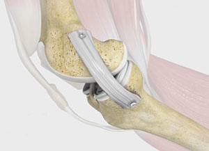
The surgical procedure for medial collateral ligament reconstruction involves the following steps:
- Your surgeon will make an incision over the medial femoral condyle.
- Care is taken to move muscles, tendons, and nerves out of the way.
- The donor tendon is usually harvested from the Achilles tendon.
- The soft tissue around the femur is debrided to assist in the insertion of the Achilles bone plug.
- For placing the graft, a tunnel is created from a guide pin to the anatomic insertion of the MCL on the tibia, using the index finger and surgical scissors.
- The Achilles tendon graft is inserted in the femoral tunnel and fixed using screws.
- The MCL graft is made taut, with the knee at 20° flexion under varus stress, and fixed to the tibia with a screw and a spiked washer.
- The incision is closed with sutures and covered with sterile dressings.
Postoperative Care following MCL Reconstruction
In the first two weeks after the surgery, toe-touch and weight-bearing are allowed with the knee brace locked in full extension. After 2 weeks, 0° to 30° of motion is allowed at the knee. At 4 weeks, knee flexion is allowed from 60° to 90° of motion and full weight-bearing is permitted. At 6 weeks, the brace is removed, and you are allowed to perform full range of motion. Crutches are often required until you regain your normal strength.
Risks and Complications of MCL Reconstruction
Knee stiffness and residual instability are the most common complications associated with MCL reconstruction. The other possible complications include:
- Numbness
- Infection
- Blood clots (deep vein thrombosis)
- Nerve and blood vessel damage
- Failure of the graft
- Loosening of the graft
- Decreased range of motion
Related Topics:
- Partial Arthroscopic Meniscectomy
- Intraarticluar Knee Injection
- Combined Hyaluronic Therapy for the Knee
- Arthroscopic Debridement
- Failed Meniscus Repair
- Meniscal Transplantation
- Meniscectomy
- Chondroplasty
- Viscosupplementation
- Physical Therapy for Knee
- Knee Arthroscopy
- Joint Preserving Osteotomy
- Multiligament Reconstruction of the Knee
- Arthroscopic Reconstruction of the Knee for Ligament Injuries
- PCL Reconstruction
- LCL Reconstruction
- ACL Reconstruction
- ACL Reconstruction of Patellar Tendon
- ACL Reconstruction Procedure of Hamstring Tendon
- Bridge Enhanced ACL Repair (BEAR)
- MCL Reconstruction
- Cartilage Replacement
- Autologous Chondrocyte Implantation
- Partial Meniscectomy
- Meniscal Surgery
- Revision ACL reconstruction
- Patellar Realignment