Meniscectomy
Meniscectomy is a surgical procedure indicated in individuals with torn meniscus where the conservative treatments fail to relieve the pain and other symptoms. Meniscectomy is recommended based on the ability of meniscus to heal, patient’s age, health status, and activity level.
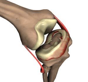
The meniscus are the two C-shaped pieces of cartilage located between the thighbone (femur) and shin bone (tibia) that act as shock absorbers and cushion the joints. The meniscus distributes the body weight uniformly across the joint and avoids the pressure on any one part of the joint and development of arthritis. Because it is affected by weight bearing, the meniscus is prone to wear and tear and meniscal tear is one of the most common knee injuries. Meniscal tears may be developed by people of all ages and is more common in individuals who play contact sports. In addition to degenerative, wear-and-tear type of meniscus tears, younger patients may sustain a meniscus tear with an acute injury.
Meniscus tears may have different appearances such as longitudinal, parrot-beak, flap, bucket handle, and complex tears. For acute tears, sudden twisting, squating, or bending are the most common causes. Meniscus tears often cause severe pain, stiffness, swelling, catching or locking of the knee, and may limit the movement. Meniscus tears are often suspected with the presenting symptoms and diagnosed with an advanced imaging study like an MRI.
Treatment
For degenerative meniscus tears in older patients, conservative treatment is often begun first and consists of R.I.C.E (Rest, Ice, Compression, and Elevation) use of non-steroidal anti-inflammatory medications, formal physical therapy, and/or corticosteroid injections. If conservative management fails to relieve the symptoms, surgery is recommended and performed using an arthroscopic technique. Depending on the extent and pattern of the tear, your surgeon will decide on whether to perform a partial meniscectomy (where the unstable meniscal fragments are removed intact tissue is left in place and the edges are smoothened) or a meniscus repair.
Arthroscopic Meniscectomy
- The arthroscope is a small fiber-optic viewing instrument made up of a tiny lens, light source, and video camera. The surgical instruments used in arthroscopic surgery are very small (only 3 or 4 mm in diameter), but appear much larger when viewed through an arthroscope.
- The television camera attached to the arthroscope displays the image of the joint on a television screen, allowing the surgeon to look throughout the knee at cartilage and ligaments, and under the kneecap.
- The surgeon makes two small incisions (about 1/4 of an inch), around the knee to enter the knee joint. Each incision is called a portal. In one portal, the arthroscope is inserted to view the knee joint. In the other, instruments are inserted to assist in the completion of the procedure.
- Along with the arthroscope, a sterile solution is pumped into the joint which expands the viewing area, giving the surgeon a clear view and room to work.
- With the images from the arthroscope as a guide, your surgeon can look at the menisci and confirm the type, location, and extent of the tear. Once your surgeon has located the meniscal tear, surgical biters and shavers are inserted into the portals to remove the torn menisci. The incisions are then closed with absorbable sutures, a bandage is applied, you are awakened from anesthesia, and brought back to the recovery room.
- In total meniscectomy, entire menisci are removed and in partial meniscectomy, only the torn part of the tissue is removed leaving the intact tissue in place with edges smoothened.
Related Topics:
- Partial Arthroscopic Meniscectomy
- Intraarticluar Knee Injection
- Combined Hyaluronic Therapy for the Knee
- Arthroscopic Debridement
- Failed Meniscus Repair
- Meniscal Transplantation
- Meniscectomy
- Chondroplasty
- Viscosupplementation
- Physical Therapy for Knee
- Knee Arthroscopy
- Joint Preserving Osteotomy
- Multiligament Reconstruction of the Knee
- Arthroscopic Reconstruction of the Knee for Ligament Injuries
- PCL Reconstruction
- LCL Reconstruction
- ACL Reconstruction
- ACL Reconstruction of Patellar Tendon
- ACL Reconstruction Procedure of Hamstring Tendon
- Bridge Enhanced ACL Repair (BEAR)
- MCL Reconstruction
- Cartilage Replacement
- Autologous Chondrocyte Implantation
- Partial Meniscectomy
- Meniscal Surgery
- Revision ACL reconstruction
- Patellar Realignment